Obstetric Brachial Plexus (OPB)
Obstetric Brachial Plexus Surgery in Madrid
There are five nerve roots (C5-T1 roots) called the brachial plexus. They connect the muscles of the shoulder, elbow, and hand to the spinal cord.
Obstetric brachial plexus (OBP) injuries occur during vaginal delivery.
Incidence varies between 0.4 and 4 per 1000 live births.
SYMPTOMS
It typically presents with flaccid paralysis of the arm. The characteristic position of the arm is the shoulder in internal rotation, elbow extended, and the forearm pronated (arm close to the body). The hand is usually positioned with a closed fist and an adducted thumb. Depending on the extent of the injury, the presentation may vary:
- C5-C6 root lesions: movement of the shoulder and flexion of the elbow are affected. The movement of the hand is preserved.
- C7-T1 root lesions: involvement of the hand and wrist. They are usually associated with more complex nerve injuries.
- Complete (C5-T1): they present involvement of the entire arm – shoulder, elbow, wrist, and hand.
TYPES OF INJURIES
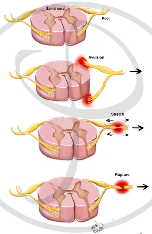
Nerve injury of obstetric brachial plexus can be caused by different mechanisms. The type of treatment will be determined by causative mechanisms. There are 3 types of injuries:
- Nerve avulsion: it is a pulling out of the nerve roots of the medulla.
- Nerve stretch: The nerve is still connected to the spinal cord and is in continuity. However, it cannot transmit information due to injury within the nerve itself.
- Nerve section (rupture): The nerve is connected to the spinal cord, but it is not in continuity (it is cut).
MECHANISMS OF NERVE ROOT INJURY
There are three types of nerve root injuries. Avulsion is the most severe injury, because it is not possible to connect the nerve to the spinal cord. The stretch and section (rupture) of the nerve are less severe.
DIAGNOSIS
The diagnosis is made based on:
- Clinical history: information told by the parents.
- Physical examination: assessment of the strength and sensation of the affected limb.
- Electrophysiological studies: they help determine the type and location of the nerve injury. They are useful to differentiate avulsion injuries vs. stretching or rupture of the nerve.
- Imaging studies: MRI should be done. Level and type of injury (avulsion vs stretching or rupture) can be distinguished.
SURGICAL TREATMENT
It is important to understand the following concepts:
WHEN TO PERFORM A SURGERY
A muscle that is not connected to the nerve atrophies over time. After 1 year the muscle cannot be recovered. On the other hand, if the nerve is in continuity (it is not cut), spontaneous recovery of the nerve and muscle is possible over time without surgery. The nerve grows at a rate of 1 mm / day, starting from the third week after the trauma.
The balance between “waiting” for spontaneous nerve recovery and “not waiting – performing surgery” (due to muscular atrophy) is complex and must be decided by a brachial plexus specialist. As a rule, surgical treatment should be carried out within 6 months after the injury.
It is very useful for parents to record (for example, with a mobile phone) the movements, to show them in the clinic to the specialist.
It is important to note that surgery of these characteristics, due to its complexity, usually requires careful planning. For this reason, it is important to consult a brachial plexus specialist as soon as possible.
AVAILABLE NERVES FOR RECONSTRUCTION
When planning a reconstruction, it is important to know which nerves are potentially donors. In a nerve avulsion, the damaged root cannot be used since it is not possible to reconnect a nerve root to the spinal cord. Therefore, it is necessary to look for nerves that supply this function. In cases where no nerve of the arm works (complete brachial plexus injuries – they are the most severe), nerves out of the brachial plexus (the spinal nerve or intercostal nerves) can be used.
PRIORITIES IN THE RECONSTRUCTION
In cases where there is a limitation of available nerves (for example, in complete brachial plexus injury), it is important to prioritize the muscles to be recovered. As a rule, the most important movements are: flexion of the elbow; external rotation or abduction of the shoulder and hand grasp.
SURGICAL TECHNIQUES
NEUROLYSIS
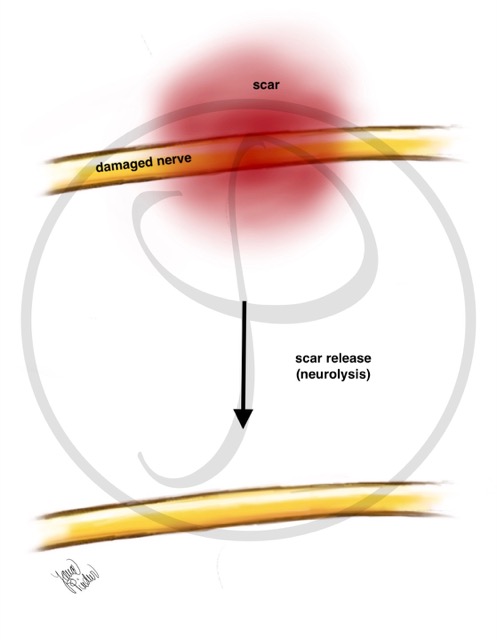
Neurolysis consists of removing all the scar that surrounds the damaged nerve. This is usually required in blunt trauma (for example, after a dislocated elbow without skin injury).
NEUROLYSIS
When a nerve is damaged, a scar appears around it that prevents its proper functioning. Neurolysis is the surgical technique that removes this scar, freeing the nerve to regenerate.
NERVE DECOMPRESSION
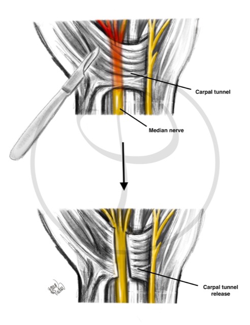
It consists of freeing the nerve from an anatomical structure (usually a ligament or tendon).
NERVE DECOMPRESSION
Releasing a nerve that is compressed by a ligament causes its recovery. This occurs in the carpal tunnel syndrome, where the median nerve is “trapped” at the level of the wrist.
NEURORRHAPHY
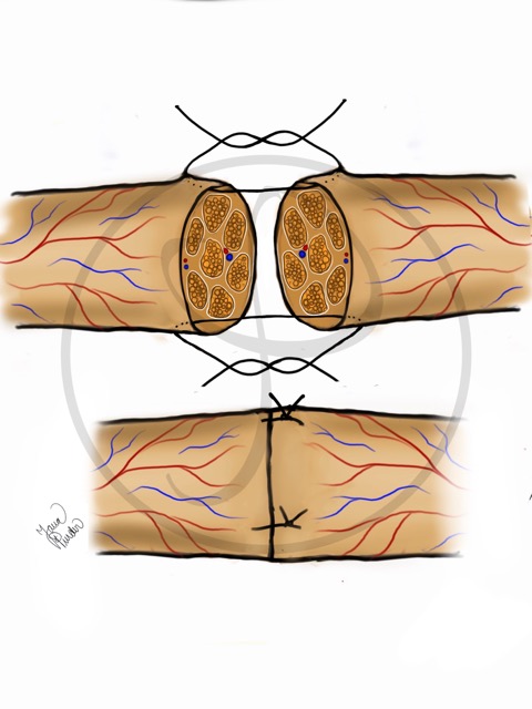
Neurorrhaphy consists of joining the ends of the divided nerve. This procedure requires a microscope and a very fine suture (thread) to join the ends of the nerve. It is required in wounds with a cut nerve.
NEURORRHAPHY
Using a very fine thread, the ends of the cut nerve are joined under a microscope.
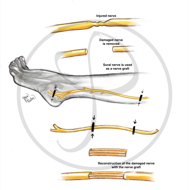
NERVE GRAFT
Nerve graft consists in obtaining a nerve from another location to repair the gap between the two ends of a cut nerve (as a ‘bridge’). This technique is used when the nerve has been cut and there is a separation between the two ends that does not allow direct neurorrhaphy (suture).
Generally, it uses the sural nerve as donor nerve. This nerve is responsible for the sensation of the external part of the foot, so its use leaves minor sequelae.
NERVE GRAFT
The sural nerve (leg nerve) can be used to bridge a nerve that has been damaged (from the brachial plexus or from any other location).
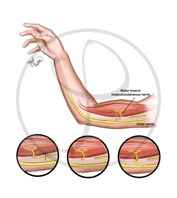
NERVE TRANSFER
Nerve transfer consists of joining a part of a healthy nerve to the damaged nerve. This technique is used when the nerve section is too far from the muscle to be reinnervated (reconnected), or in cases where the proximal end of the nerve is not available (typical in high-energy trauma, such as in motorcycle accidents over 100 km / h).
NERVE TRANSFER
It consists of connecting a part of a healthy nerve to a nerve that has been injured. In this figure, a part of the ulnar nerve (nerve without injury) is connected to the musculocutaneous nerve (previously damaged nerve).
TENDON TRANSFERS
A tendon of a muscle that works relates to another tendon of a muscle that does not work. This technique is used when nerve reconstruction has not been performed and there is muscle atrophy, for example in chronic injuries, more than one year of evolution.
TENDON TRANSFER
It consists of connecting a working tendon to another tendon that has been injured. In this figure, one of the two tendons that extends (stretches) the index finger (uninjured tendon) is connected to the tendon that moves the thumb (previously damaged tendon).
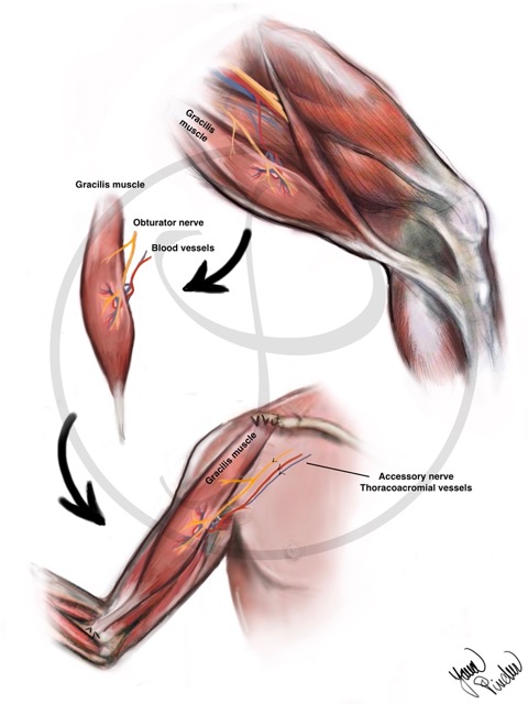
FREE MUSCLE TRANSFER
It involves transplanting a muscle from another part of the body to the damaged limb to provide missing function. This technique is used when reinnervation of the damaged muscle is not possible and there is no possibility of a tendon transfer.
FREE MUSCLE TRANSFER
It consists of transplanting a muscle from another part of the body (generally the leg) to the damaged area. It is necessary to connect the blood vessels of the muscle (they give nutrition) and a nerve.
SECONDARY SURGERIES
To improve the limb function, sometimes it is necessary to perform other interventions after the first surgery. These procedures usually consist of contracture release and tendon transfers (previously described). Such procedures are usually carried out at school or preschool age.
POSTOPERATIVE CARE
After surgery, rehabilitation plays an essential role. Success or failure of the surgery largely depends on the quantity and quality of the rehabilitation performed. There are 3 phases in the post-surgical period:
- Immobilization phase: a splint is used to immobilize the arm and avoid damage in the repaired nerves. The maximum time is usually 3 weeks.
- Passive mobilization phase: passive exercises to avoid joint stiffness. It begins after the immobilization phase until an active movement of the joint is possible.
- Active mobilization phase: Exercises to activate the target muscle. They should be started after the immobilization phase. Sometimes it takes more than 1 year to get active movement of the affected muscle. Due to the long period of this phase, a high degree of involvement and motivation is crucial.
WHAT TO DO IF YOU SUSPECT YOUR CHILD
HAS A BRACHIAL PLEXUS INJURY
- Visit a Brachial Plexus Injury Specialist – Although it is not an emergency, you should find a specialist team over the next few days or weeks.
- Rehabilitation is essential. Systematically perform the exercises recommended by your specialist. Even though the nerve regenerates, and the muscle works again, you will not be able to make movements if the joint is stiff and without mobility.

